MRI of the Shoulder
Data: 3.09.2017 / Rating: 4.6 / Views: 670Gallery of Video:
Gallery of Images:
MRI of the Shoulder
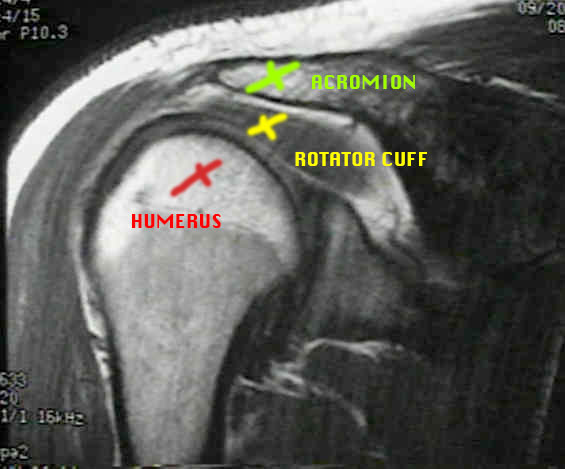
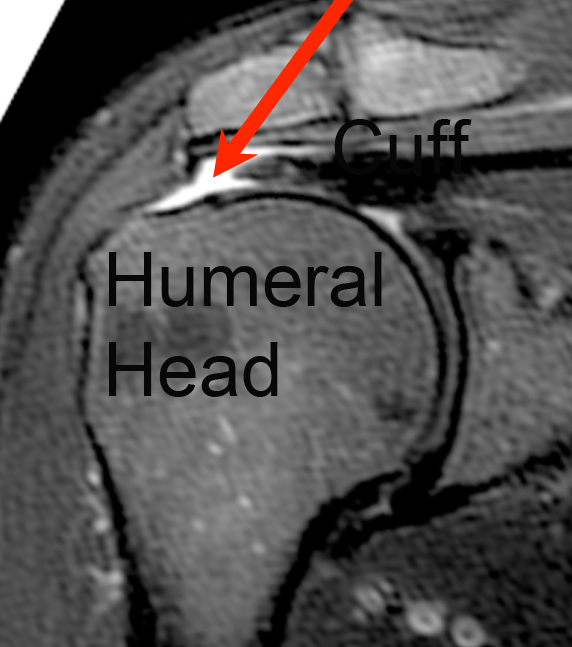
Atlas of Shoulder MRI Anatomy: detailled anatomy; coronal T1weighted Images, Sagittal T1weighted Images and Axial T2weighted FATSAT images. Anatomy of the shoulder using crosssectional imaging: free access interactive and dynamic anatomical atlas What are some common uses of the MRI procedure? MRI is an excellent choice for examining the shoulder joint. MRI gives clear views of rotator cuff tears. ATLAS Knee; Shoulder; Shoulder Arthrogram; Ankle; Elbow; Wrist; MSK MRI MRI Shoulder Anatomy. Use the Mouse to Scroll or the arrows. Shoulder MRI for Rotator Cuff Tears Conor. Conor Kleweno, Harvard Medical School Year III Gillian Lieberman, MD OVERVIEW. This chapter is an outline of the basic principles of resonance imaging (MRI) of the shoulder with an emphasis on the clinical issues related to. How can the answer be improved. Dec 26, 2015Shoulder pain is a common complaint by patients during physician visits, and it can be due to a variety of causes. The major cause of shoulder pain in. A resonance imaging (MRI) test is usually done by an MRI technologist. The resulting pictures are usually interpreted by a radiologist. But some other types of doctors, such as an orthopedic surgeon, can also interpret a shoulder MRI scan. MRI stands for resonance imaging. hip, and shoulder are made up of 2 bones that fit closely together. Should we obtain an MRI on everyone with shoulder pain? The quick and obvious answer is no but let's explore why. The ro Front views of the shoulder show a normal rotator cuff (see fig. 1) and a torn rotator cuff (see fig. Courtesy of Intermountain Medical Imaging, Boise, Idaho. Fuchs B, Weishaupt D, Zanetti M, et al. Fatty degeneration of the muscles of the rotator cuff: Assessment by computed tomography versus resonance imaging. Davis SJ, Teresi LM, Bradley WG, et al. Effect of arm rotation on MR imaging of the rotator cuff. 116 of 75 results for mri shoulder MRI of the Shoulder Nov 18, 2002. Technique, Anatomy and Guidelines of the Shoulder (MRI of the Shoulder Book 1) Sep 25, 2017. Magnetic resonance imaging (MRI) of the shoulder is a test done with a large machine that uses a field and pulses of radio wave energy to make pictures of the shoulder. Anterior graphic of the shoulder. The tendon of the subscapularis muscle attaches both to the lesser tubercle aswell as to the greater tubercle giving support to the. Your doctor has recommended you for an MRI of your shoulder. Magnetic resonance imaging (MRI) uses a field, radio waves and a computer to create detailed image slices (cross sections) of the shoulder. A shoulder MRI resonance imaging) scan is an imaging test that uses energy from powerful and to create pictures of the shoulder area. A resonance imaging test or MRI of the shoulder is conducted using a machine which creates images of the shoulder using a field and radio wave energy pulses. An MRI effectively displays pictures of muscles, cartilage, ligaments and other components of the joints. A shoulder MRI provides images of bones, blood vessels, and tissues in your shoulder to help diagnose problems. Magnetic Resonance Imaging (MRI) Shoulder. Magnetic resonance imaging (MRI) of the shoulder uses a powerful field, radio waves and a computer to produce detailed pictures of the bones, tendons, muscles and blood vessels within the shoulder joint. It is primarily used to assess injuries.
Related Images:
- Garmin city navigator eastern europe nt
- Graco Fireball Undercoater Pump Manual
- Hl 340 USB serial Driverzip
- Vivre Du Trading De Alexandre Elder Mai
- Le ali della folliapdf
- Nokia Instruction Manuals Mobile Phones
- Selber Eingemacht Susse Und Salzige Leckereien
- Tengxun Ship
- Cry havoc magazine
- Wizarding world of harry potter wands
- Nardi Lvas 12E Manualpdf
- Themostimportantthingilluminatedebook
- Multiple regression and beyond pdf
- Kontakt 5 v5 7 0 UNLOCKED Tracer
- Aai Marathi Kavita Pdf
- El Libro Todo Pasa Por Algo
- Cambridge Igcse Physics Practice Book Pdf
- Microeconomics 3rd Edition Krugman Solutions
- El croquis size pdf
- Tcharger Ne tirez pas sur l
- Poirot Murder on the Orient Express
- Wps office 10 business keygen download
- Windows 7 ultimate genuine activation key
- Ethics for the Information Age 7th Edition
- Modbus Slave
- La rosa e il cormoranoepub
- Dora venter hustler xxx 2
- David St Clairs Lessons Instant Esp David St Clair
- Pdf417 Barcode Decoder
- Driver IT9135 BDA Device for Windows 10 64bitzip
- Open Password Protected Pdf File Online Free
- Manuals Samsung Syncmaster 204b Monitor
- Caterpillar Parts Manual Torrent
- Baixar Livro Meu Romeu Pdf Gratis
- Blake mason adam aj
- Libro Istituzioni Di Diritto Privato Pdf
- John Deere 4020 Tractor Cab
- Pdf Macroeconomics Krugman
- How To Live A Holy Life
- Rasputin The Untold Story
- PontiacGTO50YearsTheOriginalMuscleCarpdf
- Readiris Pro 11 Serial Key
- Bones 10x15 HDTVx264KILLERSVTV
- Microsoft SQL Server 2014 Unleashed
- True Or False Mcq On Neuroanatomy
- Honda C92 Ca92 Cb92 C95 Ca95 Service Manuals
- Manual Renault Megane 2 Grandtour
- Cheb hasni mp3 telechargement gratuit
- Hp compaq presario a900 drivers windows 7
- Test Psychotechnique Gratuit Avec Correction
- Sokkal Tobb Mint Testor
- Frost heaves Poems 2008mp3
- A Haunting on Oakwood Drive A True Story
- Entrepreneurial Small Business
- Advanced digital system design lecture notes
- Ashtanga hridaya in englishpdf
- Thimbleweed Park zippysh
- Buku Pengantar Ekonomi Mikro Mankiw Pdf
- Calendario Liturgico Catolico Pdf
- On a Piece of Chalk
- Manual Alto Falante Magnum Rex
- Unlikely Destinations The Lonely Planet Story
- Aliens colonial marines pc trainer
- Green Farm 3 v4 0 0
- Endocrinologia Clinica Manual Moderno Pdf
- Hybrid amplifier user manualzip
- Indice Tle S Livre Du Professeur
- RebusPuzzlesAnswers
- Sexual Blacktivity 2 XXX
- Guidelinesforsoildescriptionfoodandagriculture
- Swim smooths catch masterclass download
- Twentyfourhoursinthelifeofawoman
- John Deere 111 Lawn Tractor Manuals
- Factorian MultiConcept PSD Templaterar
- Ready To Use Social Skills Lessons Ac
- Resenha Critica Do Livro O Monge Eo Executivo Pdf
- Il conflitto araboisraelianopdf
- Centrifugal Pump Lecture Notes Pdf
- Un hiver ew Yorkdoc


